Fig. 3.
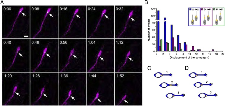
Changes in Golgi apparatus localization before and after nucleokinesis. (A) Representative time-lapse sequence showing the localization of the Golgi apparatus in migrating neurons, labeled by GalT–EGFP (green) and GAP–tdTomato (magenta). Arrows point to the Golgi apparatus (white). Elapsed time is indicated at the upper left corners of each image (h:min). (Scale bar, 10 μm.) Neurons irrelevant to our analysis are covered by black rectangles. (B) Relationship between localization of the Golgi apparatus and soma displacement of migrating interneurons between two successive movie frames. In the case shown by the blue column, the Golgi apparatus does not change its position relative to the soma of migrating neurons (S→S); in the magenta column case, the Golgi apparatus changes its relative position from the distal to the proximal portion of migrating neurons (D→P), and in the green column case, translocation from the proximal to the distal portion of migrating neurons occurs (P→D). These are schematically shown in Inset. The Golgi apparatus is shown in yellow. Data were analyzed from 386 pairs of movie frames in 39 cells from three embryos. The interval of movie frames was 8 min. Average soma displacements between two successive movie frames in each case were 3 ± 2 μm, 6 ± 4 μm, and 3 ± 2 μm (mean ± SD) for S→S, D→P, and P→D, respectively. The ratio of these events was 73% for S→S, 18% for D→P, and 9% for P→D. (C and D) Schematic representations of the relationship between the nucleus and the Golgi/centrosome complex during nucleokinesis. Translocation of the Golgi/centrosome complex occurred mostly in synchrony with that of the nucleus (D1→D2→D3), although the former preceded the nucleus in some instances (C1→C2→C3).

