Fig. 1.
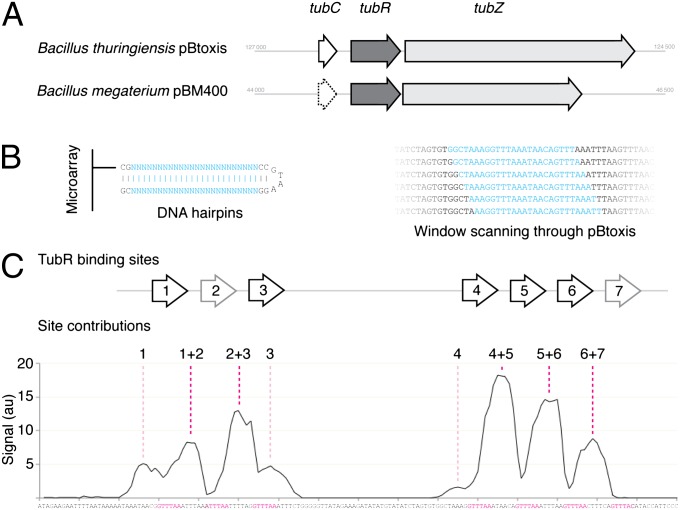
Bt tubC is composed of seven repeats, which bind TubR in a cooperative fashion. (A) Schematic comparing the tubZRC loci of Bt pBtoxis and Bm pBM400. (B) Illustration of the DNA hairpins produced on a microarray to sample the sequence of pBtoxis, and a schematic indicating how this window was scanned through a region of the plasmid sequence by successive single base pair movements. The variable window is shown in cyan, and all other base pairs in black. (C) Plot of the recorded signal for each microarray spot in a 1-bp scan over the region of Bt tubC (bp 126688 to 126496). Each point is plotted over the 12th bp of the 24-bp hairpin, with the sequence shown below. The assigned binding sites for Bt TubR are shown above, and the corresponding sequences are colored magenta below. The site(s) resulting in each peak have been annotated above the graph.

