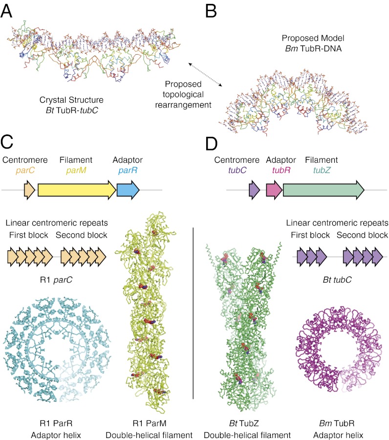
Fig. 4.
Bm TubR helix suggests further convergent evolution of type II and III partitioning systems. (A) Structure of Bt TubR (Cα ribbon representation, blue at N terminus, red at C terminus) bound to tubC (stick representation, C in white/CPK colors). (B) Structure of Bm TubR (Cα ribbon representation, blue at N terminus, red at C terminus) shown with the DNA (stick representation, C in white/CPK colors) from PDB ID code 1HW2 after superimposition of the protein chains. (C and D) Comparison of operon structure (3, 10, 12), centromeric structure (20, 21), filament superstructure (9, 14, 16), and adapter complex superstructure (18, 19, this study) for the (actin-like) ParMRC and (tubulin-like) TubZRC plasmid partitioning systems.

