Fig. 6.
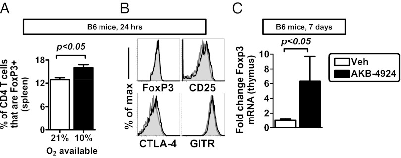
Hypoxia and HIF signaling enhance Treg abundance in vivo. (A) Whole-body hypoxia (10% O2) increases Treg abundance in the spleen after 24 h. Data show mean ± SEM, indicating percentage of live CD4 T cells that are FoxP3+, n = 4–5 mice per group, with data representative of two independent experiments and analyzed by unpaired t test. (B) Phenotype of Tregs after whole-body hypoxia, with histogram overlays comparing protein expression in Tregs in 21% (gray) or 10% O2 availability (black). (C) Treatment of mice with a PHD inhibitor increases FoxP3 mRNA expression in the thymus, measured by qPCR in thymus, in B6 mice treated with vehicle (control) or with AKB-4924. Data show mean ± SEM, n = 5 mice per group, representative of two independent experiments. Statistically significant differences are indicated, calculated by unpaired t test.

