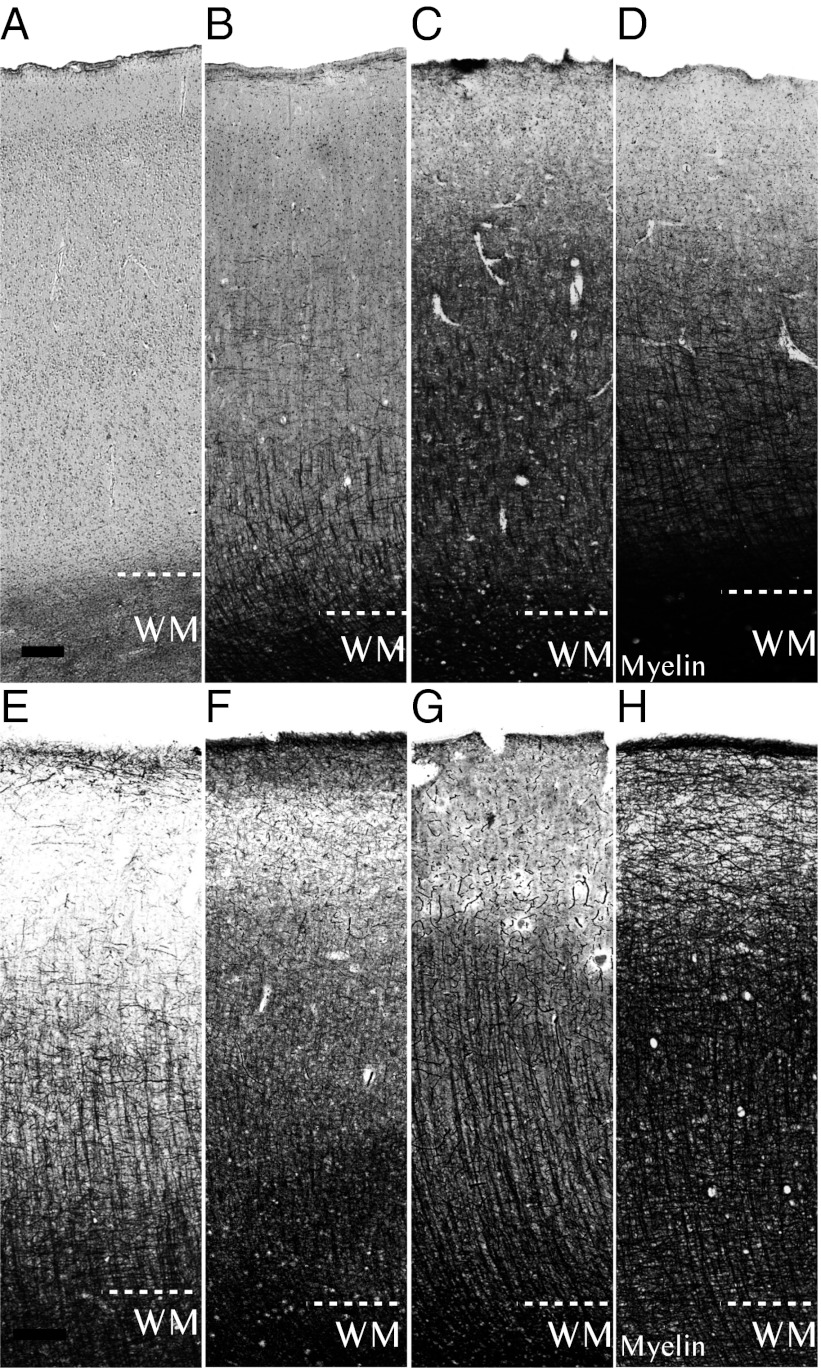
Fig. 1.
Developmental series of low-magnification photos of human and chimpanzee primary motor cortex. Sections from motor cortex (area 4) stained for myelinated axons (myelin) arranged by life-history stage. (A–D) Representative sections of human neocortical myelin as an A: infant (0–1 y), (B) child/juvenile (3–9 y), (C) adolescent/young adult (13–23 y), and (D) adult (≥28 y). (E–H) Sections of chimpanzee neocortical myelin as an E: infant (0–2 y), (F) juvenile (5–6 y), (G) adolescent (9–11 y), and (H) adult (≥17 y). White matter (WM) is demarcated at the bottom of the cortex. (Scale bar: 200 μm.)

