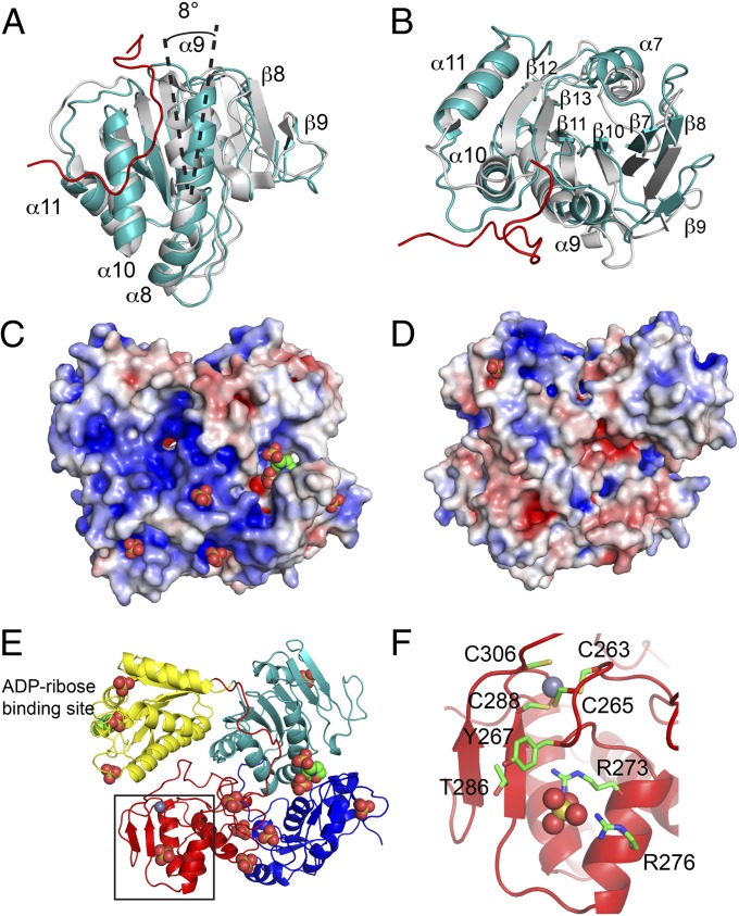
Fig. 4.
(A and B) Superposition of the MT-like domains of P23pro-zbd (teal) and nsP2pro (gray) (12). The nsP3 linker (for simplicity only residues V162-W178 are shown) connecting the macro domain and ZBD is shown in red. The view in B is rotated 90° about a horizontal axis from A. (C and D) Potential RNA-binding surface of P23pro-zbd. The location of sulfate and MES ligands are represented by spheres. Surface of P23pro-zbd is colored for electrostatic potential at ±5 kT/e. D is a 180° rotation about the vertical axis from the view in panel C. (E) Ribbon diagram of P23pro-zbd in the identical orientation as in C. (F) ZBD of nsP3 showing the four zinc-coordinating cysteines, T286, a sulfate ion represented by spheres bound to Y267, R273, and R276, and surrounding residues. The close-up view corresponds to the box in E.

