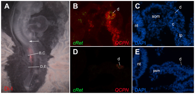
Fig. 3.
Analysis of cRet expression in transplants of prospective duct tissue into non-duct regions. Explants from stage 4 quail mid-primitive streak were stained with DiI and transplanted into the equivalent location in the primitive streak of stage 8 chick embryos, as in Fig. 2. DiI image in A indicate that the grafted cells remained below the region of the nephric duct primordia at axial level of somites 8-10 (arrow indicates somite 10). Embryos were sectioned and analyzed by combined fluorescent in situ hybridization for cRet (B,D, green) and immunofluorescence for the quail marker QCPN (B,D, red) and for DAPI (C,E). cRet-expressing graft cells are found in a morphologically normal nephric duct. c, nephrogenic cord; d, nephric duct; lp, lateral plate; nt, neural tube; psm, presomitic mesoderm; som, somite.

