
Fig. 3.
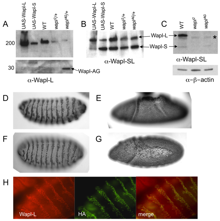
Wapl-L, Wapl-S and Wapl-AG are nuclear proteins. (A-C) Western blots using the α-Wapl-L (A) (specific for Wapl-L) and α-Wapl-SL (B,C) (the antibody reacts with both Wapl-S and Wapl-L) on embryonic (A,B) and larval (C) crude protein extracts. (A) Numbers on left show approximate molecular weights. (A,B) The identity of Wapl-L (upper band) or Wapl-S (lower band) is confirmed by overexpression of UAS-Wapl-L or UAS-Wapl-S by an arm-Gal4 driver. (A, lower panel) Western blot probed with α-Wapl-L detected an additional band in waplAG/+ embryos running about 30 kDa, close to the predicted size of a truncated Wapl-AG protein (28.6kDa). (C) An asterisk indicate the location of a crossreacting band in larval extracts (lower panel). The blot was reprobed with α-β-actin for a loading control. (D-G) Embryos stained with Wapl antibodies. Embryos overexpressing UAS-Wapl-L or UAS-Wapl-S from an engrailed-Gal4 driver (D,F) and wild-type embryos (E,G) were stained with either α-Wapl-L (D,E) or α-Wapl-SL (F,G). Engrailed-striped staining pattern shows specific nuclear staining of overexpressed Wapl proteins by the Gal4 driver. Wild-type embryos showed ubiquitous nuclear staining at all embryonic stages (except in mitotic nuclei, not shown) when stained with either α-Wapl-L or α-Wapl-SL. Embryos anterior leftwards, dorsal upwards. (D,F) Stage 15, (E) stage 8 and (G) stage 9. Native Wapl protein is present at a much lower level than the UAS-driven Wapl proteins. Pictures of wild-type embryos were taken with a longer exposure time than the UAS-Wapl embryos. (H) Wapl-AG is a nuclear protein. UAS-Wapl-AG-HA driven by engrailed-Gal4 is detected in embryos by staining with α-Wapl-L and α-HA. α-Wapl-L also reacts with endogenous Wapl-L (note red embryo in upper left corner not stained by α-HA).