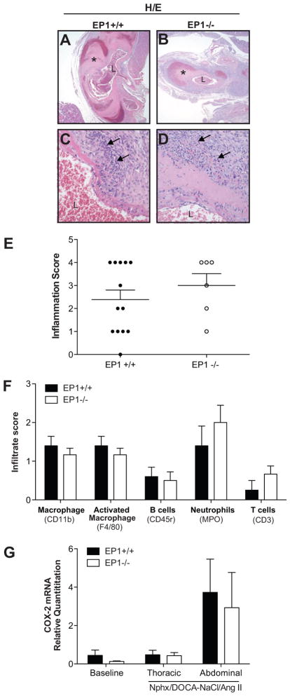
Figure 3.
Aortic inflammation. A–D. H/E stained aortae (40X and 400X magnification). A ruptured aneurysm in an EP1+/+ mouse (A, C) and a large aneurysm without rupture in an EP1−/− mouse (B, D). Vessel necrosis and perivascular inflammatory infiltration of macrophages and neutrophils are observed under high magnification (arrows, C and D). L = vessel lumen, * = aneurysm. E. Overall inflammation was scored based on H/E stained aortic sections. No differences in inflammation were observed between EP1+/+ and EP1−/− aortas (P = 0.3975, N = 5=6). F. Immunohistochemistry for detection of macrophage, B cells, T cells and neutrophils in aortae was performed and scored by in a blinded fashion by a comparative veterinary pathologist. No differences in infiltrate were observed (P > 0.05, N = 5–6). G. COX-2 mRNA in the thoracic and abdominal aortae 2–5 days post Ang II administration revealed a trend in increased COX-2 expression in the abdominal aorta of both EP1+/+ and EP1−/− mice post treatment. No significant differences were observed by treatment or between genotypes (P > 0.05, N = 3–8).

