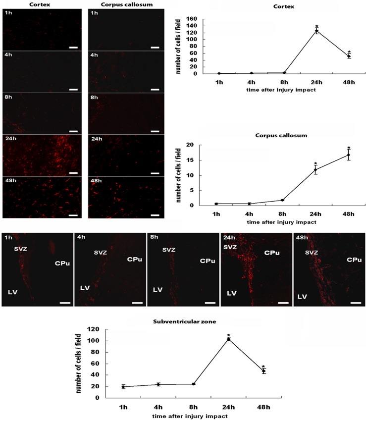
Figure 2.
Nestin expression in the cortex, the corpus callosum, and the subventricular zone after TBI. Nestin-positive cells were hardly seen in both cortex and corpus callosum at 1, 4, and 8 h post-TBI. At 24 h post-TBI, the cortex was highly populated with nestin-positive cells, and while still detected, nestin immunoreactivity was less pronounced at 48 h post-TBI. In the corpus callosum, the number of nestin-positive cells increased gradually at 24 h and peaked at 48 h post-TBI. A pattern of nestin immunoreactivity in the subventricular zone resembles that seen in the cortex. Very few nestin-positive cells were detected at 1, 4, and 8 h post-TBI, and a robust nestin immunoreactivity was recognized at 24 h post-TBI, which became less pronounced at 48 h post-TBI. Quantitative cell counts are presented in the graphs, and error bars represent mean values ± SEM. *p < 0.05 versus controls. LV, lateral ventricle; CPu, caudate putamen; SVZ, subventricular zone. Scale bars: 30 µm. Nondetectable immunofluorescence accompanied the controls.

