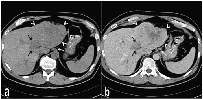
Figure 1.
Intrahepatic cholangiocarcinoma showing sarcomatous transformation in the left liver lobe. (a) On non-contrast CT, the tumour presents as a heterogeneous hypodense mass (arrow heads). (b) Portal venous phase CT reveals contrast enhancement of the tumour mass except the necrotic areas in the centre. Medial to the main tumour mass there is a more hypodense satellite tumour (black arrows), which demonstrates slight contrast enhancement on contrast-enhanced images. Note the lymph nodes at the preaortic and perigastric regions (white arrow).

