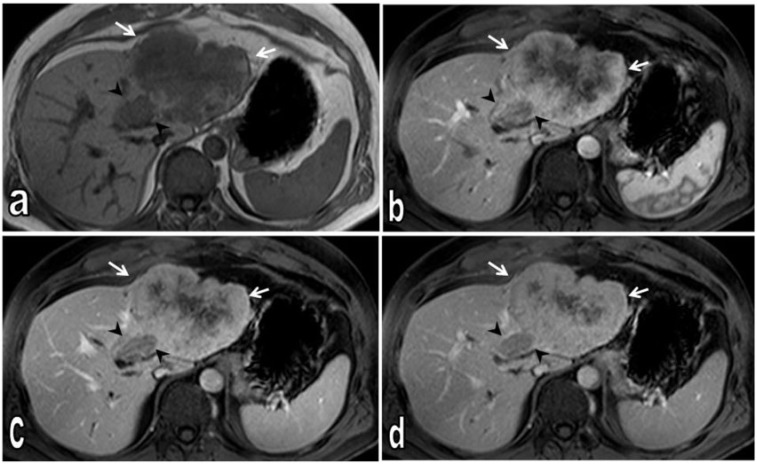
Figure 3.
Axial T1-weighted non-contrast (a), dynamic contrast-enhanced T1-weighted fat-saturated arterial (b), portal venous (c) and late venous phase (d) MR images. Non-contrast T1-weighted image demonstrates heterogeneous hypointense tumour; satellite nodule medial to the main tumour is also hypointense (arrow heads). Dynamic contrast-enhanced T1-weighted images show tumour tissue (arrows); at later phases, progressive contrast enhancement is seen from periphery to central. On late venous phases, the central region of the tumour shows no contrast enhancement consistent with necrotic changes (d). The satellite nodule shows less contrast enhancement than the main tumour on all phases (arrow heads).

