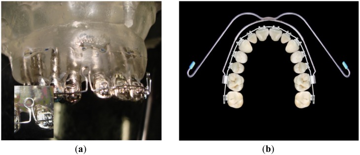
Figure 3.
(a) Picture of the maxilla model instrumented with brackets an arch wire (squared cross section) and two loop-coils; Inset: the zoom-in picture of a loop-coil opened by 3 mm for activation; (b) Occlusal image of the teeth with fixed appliance and extra-oral device connected at the first molars.

