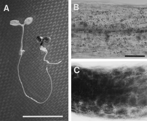
Figure 6.
Iodine staining of starch of 6-d-old wild-type and pet1-1 seedlings grown under continuous dim light in the presence of 1% Suc. A, Wild type (left) and pet1-1 (right). B and C, Magnified hypocotyl cells of wild-type and pet1-1 seedlings, respectively. Note the intensive staining of starch granules of pet1-1. Scale bars: A, 1 cm; B, 100 μm.

