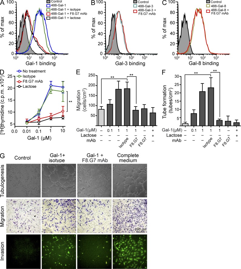
Figure 8.
A Gal-1–specific neutralizing mAb prevents Gal-1–induced EC proliferation, migration, invasion, and tube formation. (A) Binding of 20 µg/ml 488-Gal-1 to HUVEC in the presence or absence of 0.5 µM F8.G7 anti–Gal-1 mAb, 0.5 µM isotype control, or 30 mM lactose. Data are representative of three independent experiments. (B and C) Binding of 20 µg/ml 488-Gal-3 (B) or 20 µg/ml 488-Gal-8 (C) to HUVEC in the presence or absence of 0.5 µM F8.G7 anti–Gal-1 mAb. Filled histogram, nonspecific binding determined with unlabeled galectins. Data are representative of three independent experiments. (D–G) Functional activity of F8.G7 mAb in vitro. Proliferation (D), migration (E), and tube formation (F) of HUVEC incubated with or without increasing concentrations of Gal-1 in the presence or absence of 0.5 µM F8.G7 mAb, 0.5 µM isotype control, or 30 mM lactose. (G) Representative micrographs of tube formation (top), migration (middle), and invasion (bottom) of HUVEC exposed to different treatments. Data are the mean ± SEM (D–F) or are representative (G) of three independent experiments. (D–F) **, P < 0.01.

