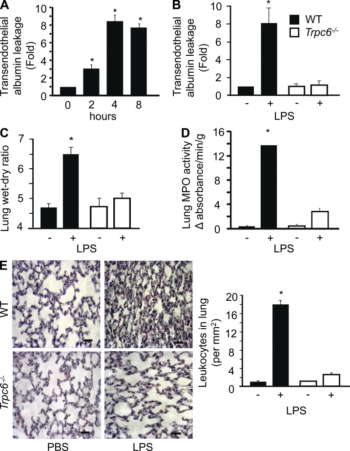
Figure 3.
Deletion of TRPC6 prevents lung vascular permeability, edema, and inflammation induced by LPS. (A) WT mice were exposed to nebulized LPS (1mg/ml) for 1 h. After 3.5 h of LPS exposure, Evans blue–labeled albumin was injected retroorbitally into each mouse to assess lung vascular hyperpermeability. Data represent mean ± SEM of four individual experiments. * indicates significant increase from the PBS-exposed group (P < 0.05). (B and C) WT and Trpc6−/− mice were exposed to nebulized LPS for 1 h. After 3.5 h of LPS exposure, Evans blue–labeled albumin was injected retroorbitally into mice. Evans blue albumin extravasation from lungs or plasma (C) and lung wet/dry ratio (D) were determined after 30 min. Data represent mean ± SEM from four individual experiments. * indicates significant increase from PBS-exposed group (P < 0.05). (D) Lungs from nebulized, LPS-exposed WT and Trpc6−/− mice were homogenized, and myeloperoxidase (MPO) activity was determined. Data are shown as mean ± SEM of three individual experiments. * indicates significant increase in MPO activity compared with WT lungs after PBS challenge or TRPC6−/− lungs receiving PBS or LPS (P < 0.05). (E) 4 h after nebulized LPS exposure, WT and Trpc6−/− lungs were fixed, sectioned, and stained with hematoxylin. The images shown at the left panel are representative sections (Bars, 10 µm), and quantitative results for leukocyte infiltration are shown in right panel. Data represent mean ± SEM of three individual experiments * indicates significant increase in neutrophils counts compared with WT lungs after PBS challenge or TRPC6−/− lungs receiving PBS or LPS (P < 0.05).

