Figure 8.
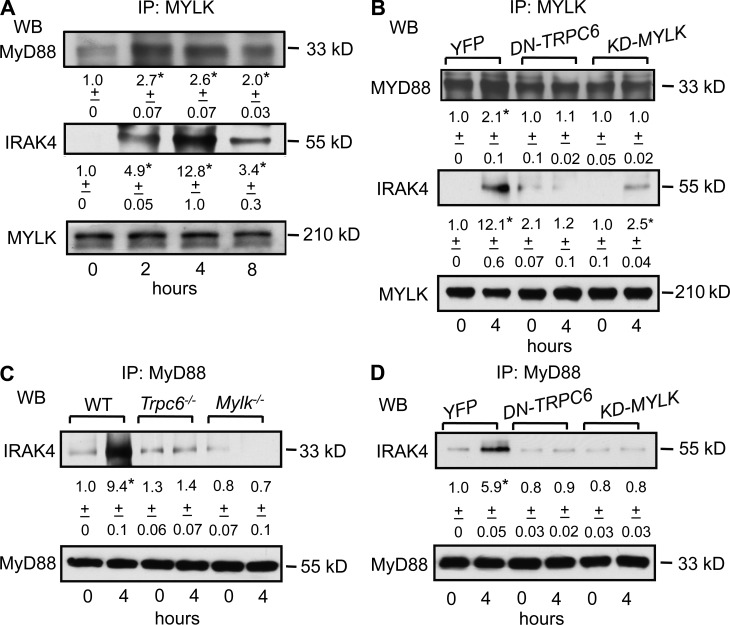
TRPC6-mediated Ca2+ entry and resultant MYLK activation induce MyD88 interaction with IRAK4. (A) WT mice were exposed to nebulized PBS or LPS for 1 h and their lungs were harvested after 2, 4, and 8 h after LPS exposure. Lung lysates were immunoprecipitated with anti-MYLK antibody, followed by immunoblotting either with anti-MyD88, anti-IRAK4, or anti-MYLK antibodies. Data represent mean ± SD of densitometric values from three individual experiments. * indicates significant increase from PBS-exposed group. (B) HPAECs were transduced either with DN-TRPC6 mutant or KD-MYLK mutant and, 24 h after transfection, cells were stimulated with 1 µg/ml LPS for 4 h. Cell lysates were either immunoprecipitated with anti-MYLK or anti-MyD88 antibody and immunoblotted with either IRAK4 antibody or MyD88 antibody to determine interaction. Data represent mean ± SD densitometric values from three individual experiments. * indicates significant increase from control transfected cells (P < 0.05). (C) Lungs from WT, Trpc6−/−, and Mylk−/− mice were harvested 4 h after PBS or 1 µg/ml LPS challenge. Lung lysates were immunoprecipitated with anti-MyD88 antibody and immunoblotted with either anti-IRAK4 or anti-MyD88 antibodies. Data represent mean ± SD of three densitometric values from three individual experiments. * indicates significant increase from PBS-exposed group (P < 0.05). (D) HPAECs transducing DN-TRPC6 mutant or KD-MYLK mutant were stimulated with 1 µg/ml LPS for 4 h. Cell lysates were either immunoprecipitated with anti-MYLK or anti-MyD88 antibody and immunoblotted with either IRAK4 antibody or MyD88 antibody to determine interaction. Data represent mean ± SD densitometric values from three individual experiments. * indicates significant increase from control transfected cells (P < 0.05).

