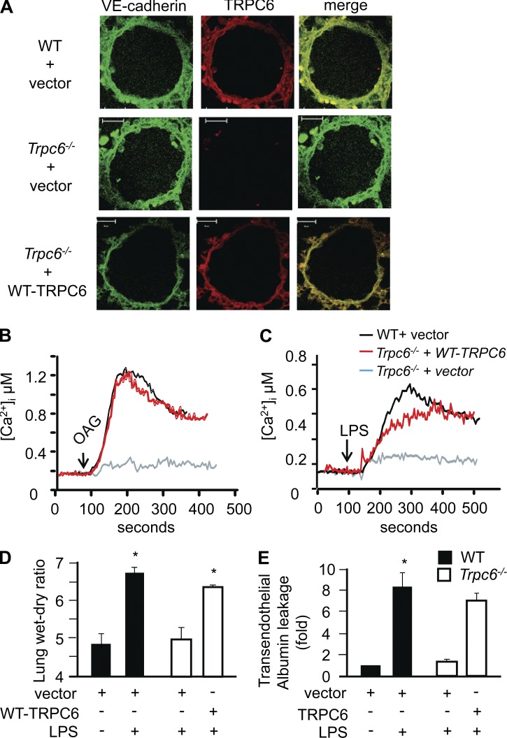
Figure 9.
Restoring TRPC expression in Trpc6−/− mice rescues LPS-induced lung vascular permeability phenotype. WT or Trpc6−/− mice were retroorbitally injected with liposome encapsulating either empty vector or WT-TRPC6 cDNA. After 48 h, lungs were harvested, sectioned and co-immunostained with anti-TRPC6 and anti-VE-cadherin antibody to confirm TRPC6 protein expression in endothelium (A). Images represent results from at least two independent experiments. Bars, 10 µm. In parallel, WT and Trpc6−/− MLECs were transfected with vector or WT-TRPC6 cDNA. After 24 h transfection, changes in intracellular Ca2+ in response to OAG (B) and LPS (C) were determined. (D and E) WT or Trpc6−/− mice expressing vector or WT-TRPC6 cDNA were exposed to nebulized 1 mg/ml LPS for 1 h. 4 h later, lung wet/dry ratio (D) and lung albumin uptake (E) were determined. Data are represented as mean ± SEM of three independent experiments. * indicates significance from WT PBS-exposed lungs or lungs not receiving LPS (P < 0.05).

