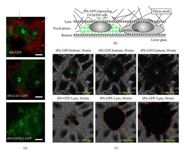
Figure 2.

Accumulation of plasminogen and effective fibrinolysis on tPA-GFP-expressing EA hy926 cells. (a) Alexa568-labeled Glu-plasminogen (568-Glu-plg) was incubated with EA.hy926 transfected with tPA-GFP, and the binding of plasminogen on the cell surface was observed (orange arrow), which was suppressed when either heavy-chain-deleted tPA-GFP (tPA(CD)-GFP) or catalytically inactive tPA-GFP (tPA(S478A)-GFP) was employed. (b) A fibrin network was formed on EA.hy926 transfected with tPA-GFP using Alexa647-labeled fibrinogen, and its spontaneous lysis was monitored by confocal microscopy. (c) Fibrinolysis initiated by tPA-GFP expressing cells, and its gradual expansion was clearly observed both focal planes of the bottom and 5 μm above the bottom. Linear binding of 568-Glu-plg was also observed at the lytic front (orange arrow), which expanded outward as the lytic area increased. A part of this figure was originally published in [4].
