Figure 3.
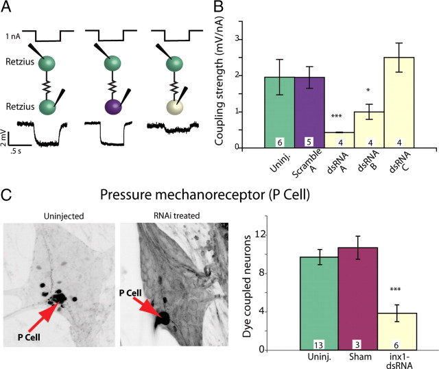
dsRNA targeting Hm–inx1 decreased function in embryonic electrical synapses. A, Current and voltage traces of embryonic Retzius-to-Retzius electrical coupling. One nanoampere of hyperpolarizing current was passed into one Retzius cell for 0.5 s, and a voltage deflection was recorded in the other Retzius cell. Green indicates an uninjected Retzius cell, purple a Retzius cell injected with scrambled dsRNA, and pale yellow a Retzius injected with active dsRNA. Injections were made at 60% ED, and the recordings were performed 48 h later, at 66% ED. B, Different dsRNA sequences produced different effects on electrical coupling. Sequences arbitrarily called dsRNAi A and dsRNAi B significantly decreased electrical coupling strength 48 h after treatment (numbers within each bar in B and C indicate the number of preparations of each type; ANOVA, *p < 0.05, ***p < 0.001; significance is compared with both uninjected and scramble-injected preparations). C, In each ganglion, a single mechanosensory P cell (red arrows) was injected with Neurobiotin; dark cells are neurons dye coupled to the Neurobiotin-injected P cell. The left image shows an untreated P cell; the right image shows cells dye coupled to the P cell 48 h after it was treated with inx1–dsRNA. The number of cells dye coupled to the P cell was significantly lower than control values for inx1–dsRNA-treated cells assayed 48 h after treatment (ANOVA, p < 0.001).

