Figure 6.

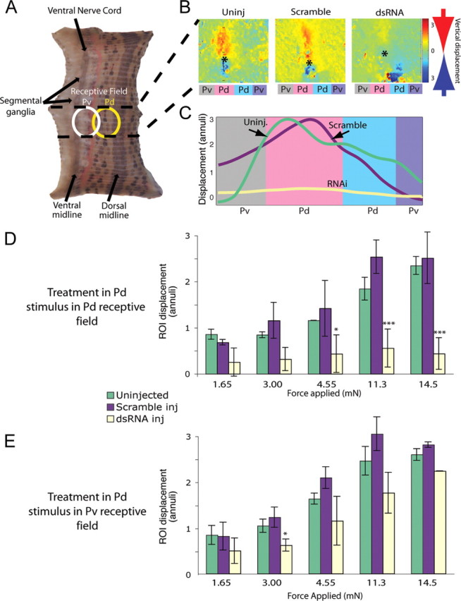
Stimulating dsRNA-treated P cells failed to elicit a behavioral response. A, Seven-segment-long piece of body wall in which only the segment containing the treated P cell remained innervated (area between the dashed lines). We applied a tactile stimulus within the receptive field of the Pd (dorsal, yellow oval) or Pv (ventral, white oval). B, Maximal longitudinal contraction after a stimulus in the receptive field of a Pd neuron. Using an optic flow algorithm (Baca et al., 2005), digital movies of contractions were converted to color-coded vector maps. Stimuli occurred at the asterisks; red indicates motion downward toward the point of touch, and blue indicates motion upward toward the touch. In both uninjected and scramble-injected controls, longitudinal movement is centered on the asterisk, indicating that the longitudinal contraction was focused on the stimulus. Colored rectangles below each image indicate the quadrant of the body wall innervated by each P cell: Pd indicates a dorsal quadrant, and Pv indicates a ventral quadrant on each side. C, Longitudinal displacement in body-wall preparations after three different treatments: uninjected (green line), Pd injected with scrambled dsRNA (purple line), and Pd injected with inx1–dsRNA (pale yellow line). Amplitudes of movements within the region of interest (ROI), normalized to the anteroposterior dimension of an annulus, were integrated to yield a local bend profile (Baca et al., 2005). D, Maximum displacement of the body-wall region of interest within the stimulated dorsal quadrant (contraction strength) as force increased (ANOVA with Bonferroni's post hoc analysis, *p < 0.05, ***p < 0.001). E, Ventral local bends in response to stimulating the receptive field of a Pv. Colors (which indicate how the ipsilateral Pd in the ganglion was treated) are the same as in D; yellow bars show results of ventral stimulation in segments in which a Pd cell was injected with im1–dsRNA (ANOVA with Bonferroni's post hoc analysis, *p < 0.05, ***p < 0.001).
