
Figure 2.
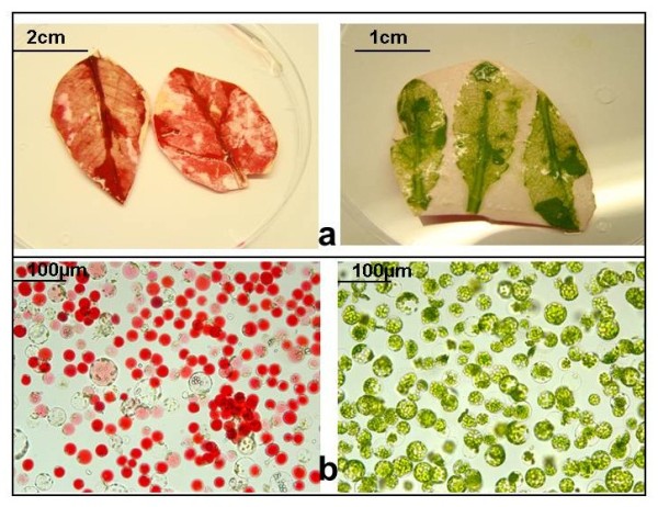
Isolation of mesophyll protoplasts from red leaves of Poinsettia (left) and Arabidopsis thaliana (right). a) Lower leaf surface after protoplast release through cellulose digestion. b) released protoplasts. Poinsettia protoplasts have large anthocyan-filled vacuoles, whereas Arabidopsis protoplasts contain numerous chloroplasts.