
Figure 10.
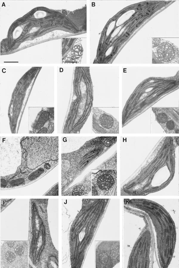
Ultrastructure of plastids from each of the cue mutants at stages of intermediate greening and at different stages for cue6. Mitochondria shown for reference in each case (inset, except for cue6). A, Wild type; B, hy1-6.2; C, phyB-17.6; D, cue3, from young leaf, toward midvein; E, cue4; F, cue6, proplastids from margin of young leaf; G, cue6, from young leaf toward the midvein (intermediate greening); H, cue6, chloroplast from mature leaf; I, cue8, from young leaf, toward midvein; J, cue9, from mesophyll cell not close to the midvein; K, cue9, from cell close to central vascular bundle. Bar in A = 1 μm; all panels are to same scale. Bar in A, inset = 0.5 μm; all insets are to same scale.