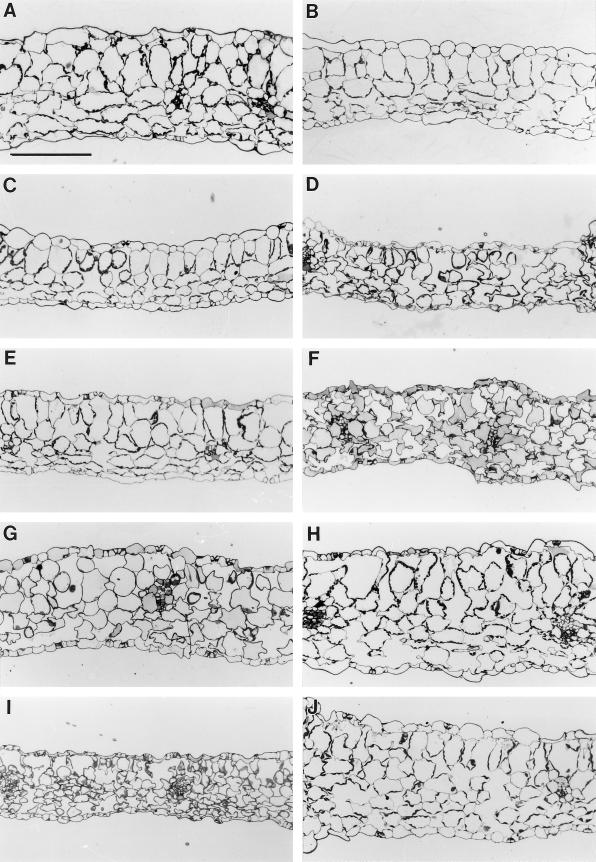
Figure 9.
Light microscopy of leaf sections of the different cue mutants. A, Wild type; B, hy1-6.2; C, phyB-17.6; D, cue3; E, cue4; F, cue6 young leaf, toward the margin (mostly pale tissue); G, cue6 young leaf, toward the midvein (increased greening); H, cue6 mature leaf (greenest tissue for this mutant); I, cue8; J, cue9 (section bordering the midvein, to the left). Bar in A = 100 μm; all panels are to same scale.

