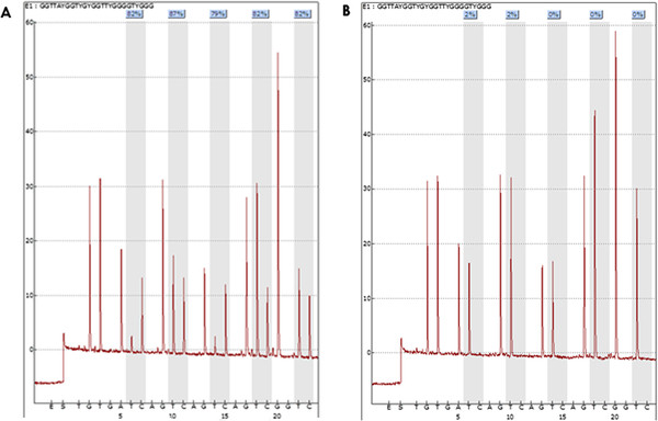
Figure 1.
Representative pyrograms with PyroMarkQ96 showing percentage of methylation at each of five CpG sites evaluated.A) Highly methylated tumor sample with an average methylation percentage of (82% + 87% + 79% + 82% + 82%)/5 = 82.4%. B) Corresponding non-methylated non-tumoral tissue from the same patient as in A with the following methylation pattern: 2%, 2%, 0%, 0%, and 0%.

