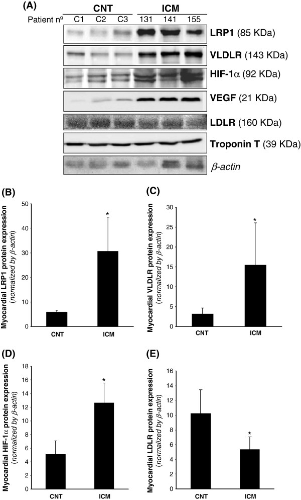
Figure 2.
LRP1, VLDLR and LDLR protein levels in control and ICM hearts. Representative Western blot analysis (A) showing LRP1, VLDLR, HIF-1α, VEGF, LDLR and troponin T protein expression in three controls and three ICM patients. Bar graphs showing the mean ± SD of protein LRP1 (B), VLDLR (C), HIF-1α (D) and LDLR (E) band quantification. Unchanged levels of β-actin were shown as loading control and used to normalize protein bands. *P < 0.05 versus CNT. CNT, controls; ICM, ischemic cardiomyopathy patients.

