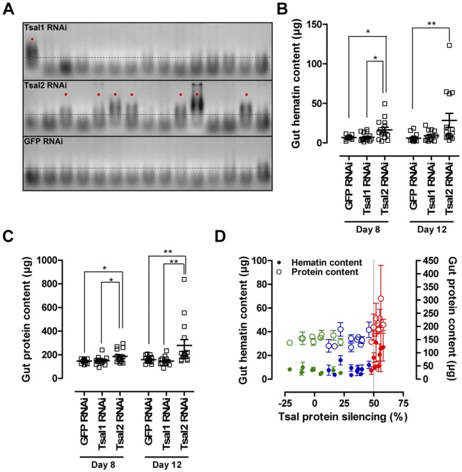
Figure 9. Effect of Tsal1 and Tsal2 specific RNAi on the tsetse fly blood meal digestion physiology.
(A) Gut homogenates of individual flies after 72 h starvation were run on a 2% agarose gel, allowing the visualization with ethidium bromide of nucleic acid degradation in the digested blood. Presented data in this panel are those for flies at 8 days p.i. A threshold of 300 bp (dotted line) was set to distinguish between the profiles of a perturbed (indicated with dots) and normal digestion. RNAi induced alterations in (B) the gut hematin digestion and (C) remainder gut protein contents 72 hours after the last blood meal. (D) Illustration of the RNAi-mediated effects on gut hematin (•, left Y-axis) and protein contents (○, right Y-axis) in function of protein silencing efficiency (Tsal1 RNAi: blue; Tsal2 RNAi: red, GFP RNAi: green). This graph represents the percentage Tsal protein reduction in the saliva harvested from 3 gland pairs (X-axis) and the average of the individually determined hematin and protein contents in the guts of the 3 corresponding flies. Phenotypic effects of Tsal2 RNAi were confirmed in 3 independent experiments. Statistical analyses were performed using the Mann Whitney test in the GraphPad Prism 5.0 software package; * and ** indicate respectively P<0.05 and P<0.01.

