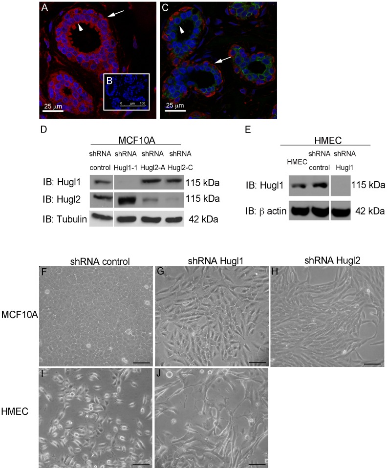
Figure 1. Hugl1 and Hugl2 display cell type restricted expression and inhibit mesenchymal phenotype.
Ductal epithelium from human breast was incubated with antibodies to either (A) Hugl1 (red), or (C) Hugl2 (red) and Keratin 18 (green), or (B) no primary antibody, secondary only control. All sections were incubated with DAPI (blue). Arrowheads indicate luminal epithelium and arrows indicate myoepithelium. Stable knockdown was established in MCF10As and HMECs with transduction of (D and E) Hugl1, (H) Hugl2 or control shRNA lentiviral particles and selected with puromycin. Protein lysates were isolated from cell lines, 20 µg of protein were separated by SDS PAGE and analyzed by immunoblot using the antibodies: anti-Hugl1, anti-Hugl2, anti-β actin (loading control), and anti-α tubulin (loading control). Molecular weights are shown at right. MCF10A control shRNA cells (F) and HMEC control shRNA cells (I) retain parental cobblestone phenotype while Hugl1 shRNA cells (G and J) and shRNA Hugl2 cells (H) take on a mesenchymal phenotype on plastic after transduction. Scale bar = 300 µm.

