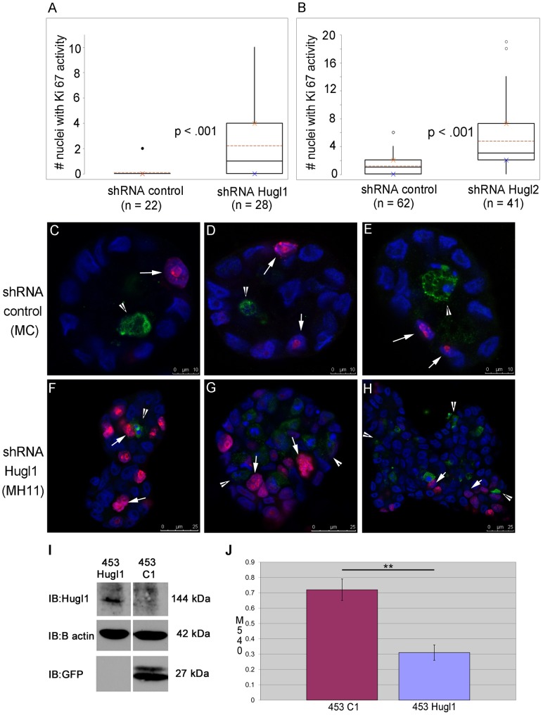
Figure 4. Hugl1 inhibits proliferation in both immortalized and transformed mammary epithelium.
Cells with stable expression of Hugl1 shRNA (MH11), Hugl2 shRNA (MH2C) and control shRNA (MC) were grown in Matrigel for 8 days, fixed with 2% PFA, permeabilized, and incubated with anti-Ki 67 (proliferation) and/or anti- cleaved caspase 3 (apoptosis) antibodies. Cells were then incubated with secondary antibodies conjugated to fluorophores (Alexa 488, 647). Acini were imaged with a Leica confocal microscope to obtain cross-sectional images from the equatorial section at 630X. (A–B) Number of nuclei with Ki-67 activity was counted per acinus. For each experiment, boxplots reflect one of three trials producing similar statistically significant results. (C–H) Acini were immunofluorescently labeled for Ki-67 (fuschia, arrows) and cleaved caspase 3 activity (green, arrowheads). Scale bars indicate size in microns. (I) Re-expression of Hugl1 in breast cancer cell line MDA-MB-453 was established with stable transfection of a fusion protein construct, pEGFP-Hugl1 (453Hugl1). Control lines were transfected with empty vector pEGFP-C1 (453C1). 20 µg of protein lysate was separated by SDS PAGE and analyzed by immunoblot, probing for either anti-Hugl1 (top panel), anti-β actin (middle panel) or anti-GFP (bottom panel). Note that EGFP-Hugl1 is a fusion protein and is 144 KDa. Molecular weight is shown at right. (J) 453C1 and 453Hugl1 were grown for 3 days and analyzed by (3-(4,5-Dimethylthiazol-2-yl)-2,5-diphenyltetrazolium bromide (MTT) assay. Statistics were performed with two-tailed Student’s t test. ** p value <0.01.

