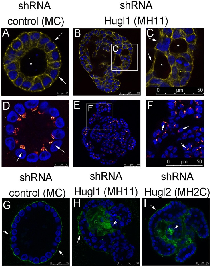
Figure 5. Hugl regulates apicobasal polarity and lumen formation in mammary acini.
Cells with stable expression of Hugl1 shRNA (MH11), Hugl2 shRNA (MH2C) and control shRNA (MC) were cultured in Matrigel for 21 days, fixed with 2% PFA, permeabilized, and incubated with primary antibodies marking apical (anti-GM130, red), cytoskeletal (anti-phalloidin, yellow), or basal (anti-Laminin V, green) domains followed by fluorescently labeled secondary antibodies (FITC, Alexa 488, or Alexa 647). Slides were mounted with antifade mounting media containing DAPI. Acini were imaged with a Leica confocal microscope to obtain equatorial cross sectional images of their morphology. (A–I) All images were taken at 630X using internal zoom. Subsections of B and E (white boxes) magnified in C and F. Scale bars indicate size in microns.

