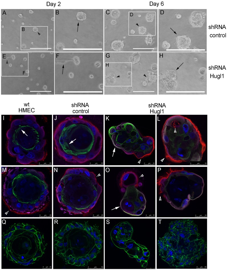
Figure 6. Hugl1 regulates membrane domain formation in HMECs.
Control shRNA and Hugl1 shRNA HMECs were cultured in Matrigel for 6 days and imaged by bright field at 20X at day 2 and day 6 of growth. White boxes in A, C, E, and G outline areas magnified in B, D, F, and H. Wild type, control shRNA, and Hugl1 shRNA HMECs (I–T) were cultured in Matrigel for 6–10 days, fixed in 2% PFA, permeabilized, and incubated with primary antibodies to apical (anti-MUC1, green, I–P), basolateral (anti-Integrin α6, purple, I–P) and basal (anti-Laminin V, red, I–P) or cytoskeletal (anti-phalloidin, green, Q–T) domains followed by fluorescently labeled secondary antibodies (FITC, Alexa 488, 594, 647). Slides were mounted with antifade mounting media containing DAPI. Acini were imaged with a Leica confocal microscope to obtain equatorial cross-sectional images of their morphology at 630X. Arrows in I, J, K and O indicate MUC 1 localization. Arrowheads in K, L, M, N, O, and P indicate Laminin V localization. Scale bars indicate size in microns.

