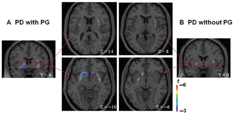
Figure 2.

Coronal and axial sections of the statistical parametric map of the change in [11C] raclopride binding potential overlaid upon the average MRI in stereotaxic space. The figure displays the significant areas of striatal dopamine release during gambling as compared to control task in (A) Parkinson’s disease patients with pathological gambling (Y = 4; Z = −10; Z = 14) and (B) without pathological gambling (Y = 0; Z = −6; Z = 8) in the ventral striatum (Z = −10 and Z = −s6) (bottom figure) and dorsal striatum (Z = 14 and Z = 8) (top figure).
