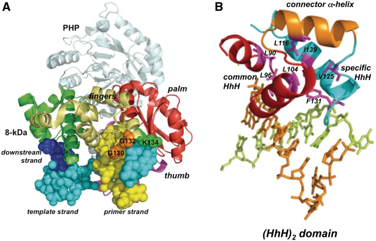
Figure 7.
(A) Ribbon representation of the structural model of PolXBs bound to DNA. Model for PolXBs was provided by the homology-modelling server SWISS-MODEL, using the crystallographic structure of the ternary complex of ttPolX as template (PDB code 3AUO) (33). The PHP domain is shown in light blue, the 8-kDa domain in green, fingers in gold yellow, palm in red and thumb in magenta. Bound DNA is represented as spheres. The upstream, template and downstream strands are coloured in yellow, cyan and dark blue, respectively. Glycines and lysine of the specifically conserved HhH motif are represented as orange and green spheres, respectively. (B) Fingers subdomain of bacterial PolXs contains a (HhH)2 domain. PolXBs structure was modelled as described in (A). Common and specifically conserved HhH motifs are represented as red and cyan ribbons, respectively, whereas connector α-helix is coloured in orange. PolXBs residues forming the conserved hydrophobic core are represented as magenta sticks. Figure was made using PyMOL software (http://www.pymol.org).

