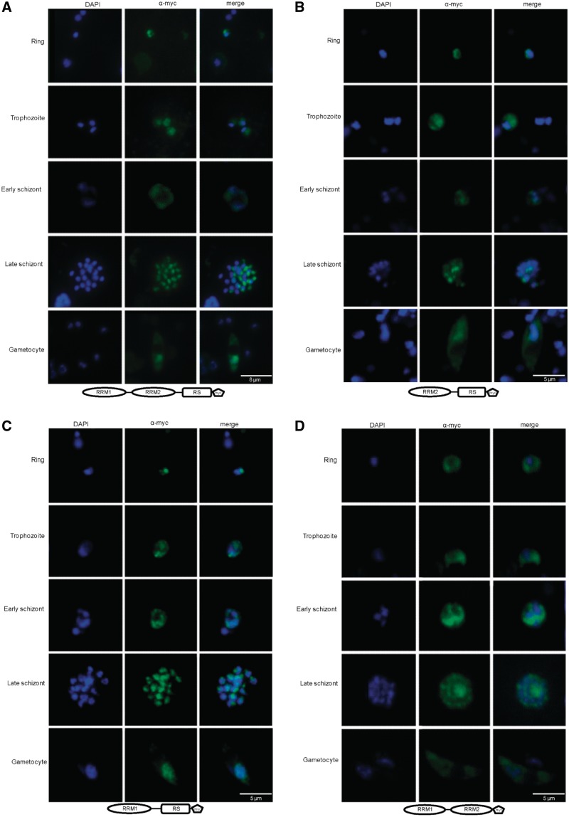
Figure 4.
The RS domain localizes PfSR1 to the nucleus. IFAs showing the localization of the entire PfSR1 as well as different PfSR1 mutants lacking either the RRM or the RS domains (ΔRRM1, ΔRRM2 and ΔRS, respectively) during IDC. The different forms of PfSR1 were ectopically express fused to a myc epitope tag and detected using anti-myc antibodies. PfSR1 (green); DAPI staining (blue); developmental stages are indicated on the left. (A) Cellular localization of PfSR1-myc. (B) Cellular localization of the PfSR1ΔRRM1-myc. (C) Cellular localization of the PfSR1ΔRRM2-myc. (D) Cellular localization of the PfSR1ΔRS-myc. The entire PfSR1 and two ΔRRM mutants shuttle between the nucleus and cytoplasm, whereas the ΔRS mutant localizes to the cytoplasm.

