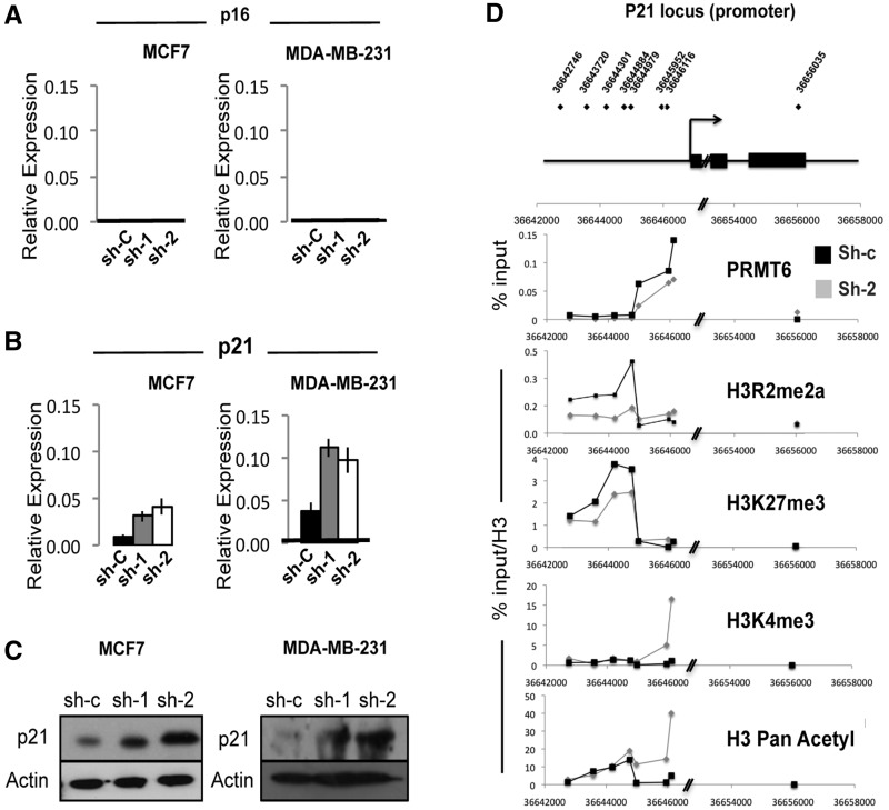
Figure 3.
PRMT6 directly represses the p21 promoter. qPCR analysis of cyclin inhibitors (A) p16 and (B) p21 in PRMT6 depleted (sh-1 and sh-2) or control (sh-c) MCF7 and MDA-MB-231 cells. (C) Immunoblot analysis of p21 protein levels in PRMT6 depleted (sh-1 and sh-2) or control (sh-c) MCF7 and MDA-MB-231 cells. (D) Chromatin immunoprecipitation in MCF7 cells using the antibody indicated on each panel. Data are shown as the % of input DNA for PRMT6 and as % of input DNA normalized to total H3 (% input/H3) for the histone PTMs. The p21 promoter in MCF7 cells is enriched for inactive- (H3R2me2a and H3K27me3) and depleted for active- (H3K4me3 and H3acetyl) chromatin marks in control cells as compared with PRMT6 depleted ones (sh-2, grey lines). Experiment was repeated four times, and a representative plot is shown.

