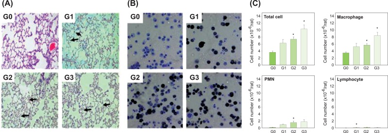
Figure 3.
Histopathological findings in the lungs and leukocytes in BAL fluid of male Sprague-Dawley rats exposed to carbon black for 13 weeks. (A) Histopathological findings. (B) Cytospun Leukocytes in BAL fluid. (C) Numbers of leukocytes in the BAL fluid. Arrows show masses of carbon black in the lungs (20x magnification). Error bars indicate the standard error of the mean. Statistically significant significances were determined by ANOVA followed by Duncan's post hoc test (p<0.05). G0, control; G1, carbon black dispersed by vortex; G2, carbon black dispersed by vortex+sonication; G3, carbon black dispersed and stabilized with albumin. BAL, bronchoalveolar lavage; PMN, polymorphonuclear leukocytes. *Statistically significant compared to control.

