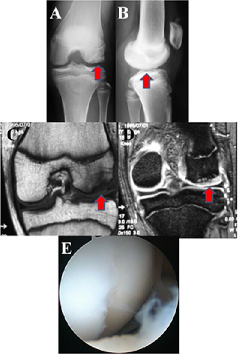
Fig. 1.
A 13-year-old girl with JOCD of the lateral femoral condyle who became asymptomatic after OAT (case 9). Anteroposterior radiograph reveals a lucent lesion (arrow) on the lateral femoral condyle (a). Lateral view (b). Coronal T1-weighted (c) and T2-weighted (d) MRI examinations of the knee performed before surgery showing a bone fragment in situ and high-signal intensity line between the bone and the fragment (grade 3) (arrow). Arthroscopic examination of the JOCD lesion of this case demonstrated a cartilage fissure in the lateral femoral condyle. The lesion was relatively stable on probing (e)

