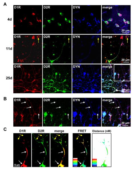
Fig. 2.
Neuronal growth and differentiation of rat neonatal striatal D1 and D2 receptor coexpressing neurons. (A) Confocal images showing D1 receptor (D1R), D2 receptor (D2R) and dynorphin (DYN) expression in rat striatal neurons following 4, 11 or 25 days in culture. Neurons showed a progressive increase in dendritic sprouting and growth after 4 days in culture. D1R and D2R showed significant colocalization with DYN in neuronal cell bodies at all time points, and in the dendrites on days 11 and 25. On day 25 of culture, some D1R and D2R coexpressing neurons began to show differential segregation of D1R and D2R to the distal dendrites (box). Neurons showing expression of only one dopamine receptor were in evidence but scarce (yellow arrows) (B) Magnification of the boxed area in (a) depicting neuronal dendrites of a D1R and D2R coexpressing neuron on day 25 of culture. While D1R and DYN exhibited colocalization in these dendrites, D2R was not present (white arrows). (C) Example of a representative neuron showing colocalization and interaction (FRET) of D1R and D2R, and the distance between the two receptors in a striatal cultured neuron. FRET between the D1R and D2R was evident in regions of receptor colocalization (white arrows) but was not present in regions absent of D1R expression (yellow arrows). Scale panel denotes FRET efficiency or distance (nM) accordingly.

