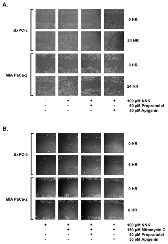
Figure 5.
Apigenin Suppresses NNK-enhanced Proliferation/Migration
BxPC-3 and MIA PaCa-2 cells were propagated to confluence in 35-mm dishes for 5-7 days. At confluence, scratch assays were performed using a 10-μL pipette tip and vacuum aspiration. Remaining adherent cells were washed, replenished with serum deficient media and allowed to stabilize overnight. (A) Cells were treated on the second day with 0.1 % DMSO vehicle control, 100 μM of NNK, 100 μM NNK with 50 μM of propranolol or 100 μM of NNK with 50 μM of apigenin for 24 hrs. Gap closures in marked areas were recorded at 10X magnification. (B) Cells were treated on the second day with 100 μM of NNK, 100 μM NNK and 150 μM of mitomycin C, 100 μM NNK and 150 μM of mitomycin C with without 50 μM of propranolol or 100 μM NNK and 150 μM of mitomycin C with 50 μM of apigenin for 6 hrs. Cell migration were observed in marked areas at 10X magnification.

