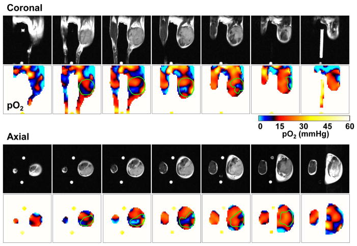
Figure 2.
Three-dimensional oxygen maps in a SCC tumor from EPRI. Oximetric images of a mouse bearing SCC tumor were acquired by EPRI at 10 mT. Sequentially, anatomic images were obtained by T2-weighted 7T MRI. Both MRI and EPRI scans were performed using an identical coil operating at 300 MHz without removing the mouse from coil assembly. The coronal and axial sliced images were serially displayed at every 2 mm.

