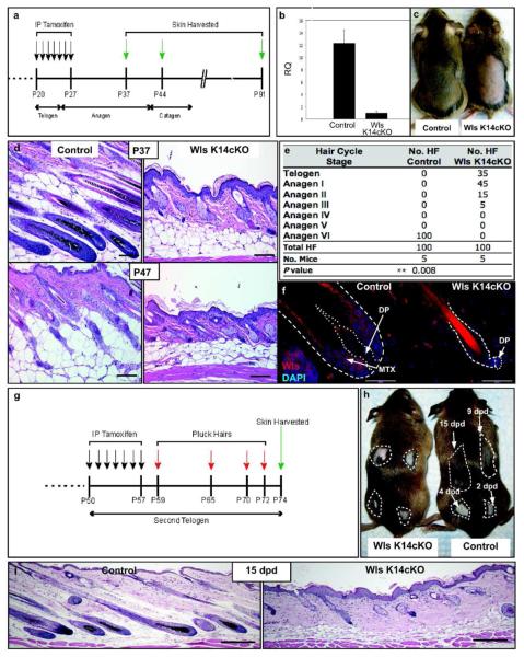
Figure 2.
Epidermal Wls is required for anagen. (a) Tamoxifen-mediated Cre induction regimen. (b) Relative quantities of Wls mRNA determined by qPCR from RNA isolated from dorsal skin epidermis of control and Wls K14cKO mice 5 days after induction (P32, N=5 mice). (c) Images of P37 mice shaved after induction. (d) H&E sections from control mice during anagen (P37) and catagen (P47; bar=100 μm). Wls K14cKO hair follicles at the same time points remained arrested in telogen or anagen I/II. (e) Hair cycle distribution of control and mutant mice at P37-40. (f) Wls expression in P37 control and mutant hair follicles (bar=50 μm). Scattered Wls immunoreactive cells were noted throughout the dermis but similar between control and mutant mice. (g,h) Tamoxifen was administered during second telogen prior to depilation at indicated times. (i) H&E sections from skin plucked 15 days post-depilation (15 dpd; bar=200 μm).

