Figure 3.
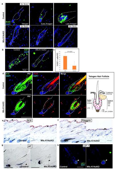
Epidermal Wnts are required for HFSC proliferation but not HFSC maintenance. (a) P37 Wls K14cKO and depilated control skin were harvested 2 hr or 6 hr after BrdU administration. Virtually no BrdU+ cells were detected immunohistochemically in the bulge/sHG of Wls K14 cKO mice even after 6hr pulse. (b,c) Ki67 immunohistochemistry of depilation-induced stage-matched control follicles compared to Wls K14cKO follicles. Graph represents results from 3 mice per group. (d) Double immunofluorescent detection of HFSC markers, CD34 and K15, in control (top panels) and Wls K14cKO (bottom) mice at P91. Illustration (right) of marker expression in normal telogen skin. (e) Immunohistochemistry of interfollicular epidermal differentiation markers K10 and (f) filaggrin (black arrows; bar=100Om). (g) AP and (h) LEF-1 expression are maintained in the DP of mutant hair follicles (arrows). Black bars=100μm, white bars=50μm.

