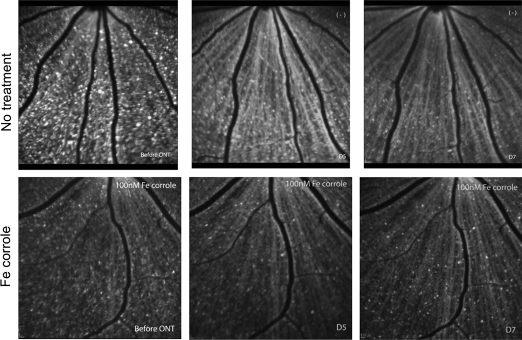
Fig. 4.
Examples of longitudinally collected images of RGCs retrograde labeled with 4Di-10Asp and detected by confocal scanning laser ophthalmoscopy. Left panels, before optic nerve transection. Middle panels, 5 days after optic nerve transection. Right panels, 7 days after optic nerve transection. The same fields are imaged in the top row or bottom row.

