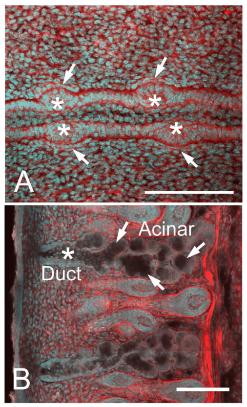
Figure 1.
Confocal images using actin (red) and nuclei (DAPI, cyan) staining of eyelids at different time points of meibomian gland morphogenesis. At E18.5 (A), we observed the formation of an epithelial condensation (asterisk) within the fused lid margin. Epithelial placodes in the superior and inferior lids also appeared opposite to each other, and were associated with condensation of mesenchyme as detected by cell alignment and increased actin staining of mesenchyme directly adjacent to the placode (arrows). Extension of epithelium into the lid mesenchyme was observed from postnatal day 0 (P0) to P8 (B) showing extensive branching to form individual acinar tissue (arrows) and the central duct (asterisk). Bar = 250 micron. (Adapted from Nien et al.8)

