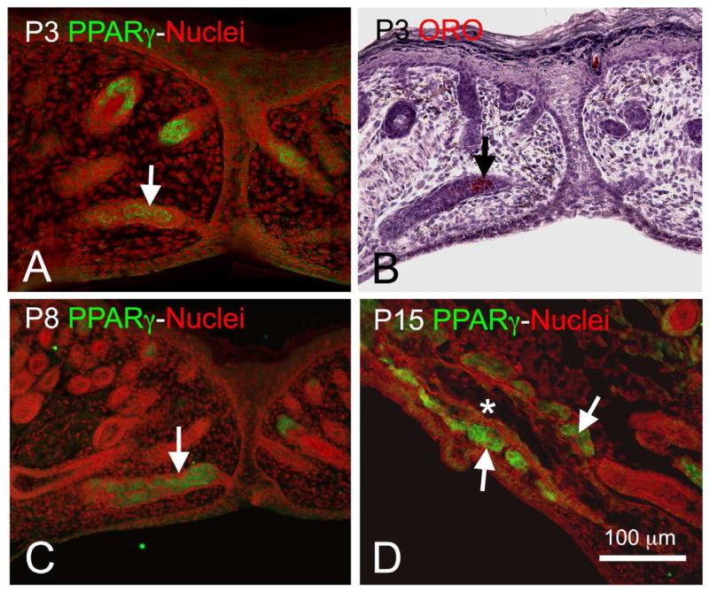
Figure 2.
PPARγ expression (Green = PPARγ, Red = nuclei) at P3 (A) was localized to the central portion of the invaginating epithelial cord (arrow) and correlated with oil-red-O staining (B, Red) indicating the presence of neutral lipid. PPARγ expression at P8 (C) was also localized to the epithelial cord (arrow) and developing acini, while at P15 (D) after eyelid opening expression was absent in the central duct (asterisk) but present in the acini (arrows). (Adapted from Nien et al.8)

