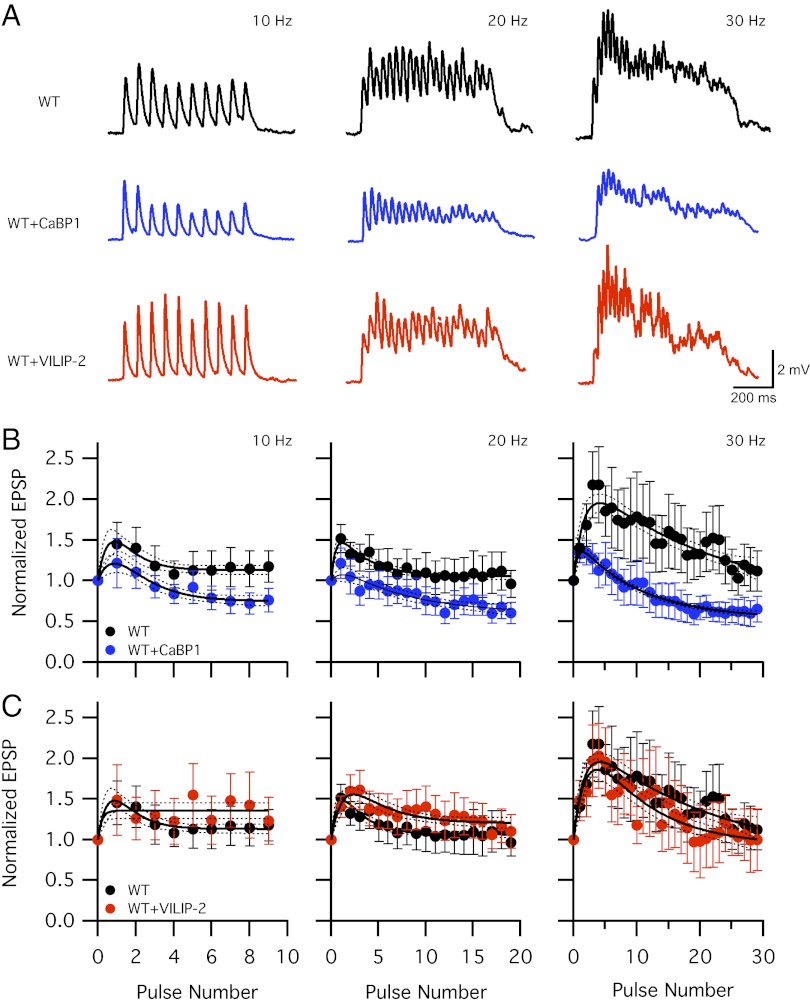
Fig. 2.
CaBP1 blocks synaptic facilitation in trains of stimuli. (A) Representative EPSPs in 1 mM extracellular Ca2+ evoked by repetitive action potentials at 10, 20, and 30 Hz for 1s in the presynaptic neurons expressing CaV2.1 alone (WT, black) or cotransfected with CaBP1 (WT+CaBP1, blue) or VILIP-2 (WT+VILIP-2, red). Data from 10 sweeps repeated every 30 s at each frequency were averaged. (B and C) Mean normalized EPSP amplitude from 10–16 synaptic pairs at 10-, 20-, and 30-Hz frequency. EPSP amplitudes were normalized to the first EPSP of each train and plotted against action potential number for WT (●), WT+CaBP1 ( ), and WT+VILIP-2 (
 ) neurons. Points represent the mean ± SEM. Solid lines are best fits of a biexponential equation as described in SI Materials and Methods. Dotted lines indicate 95% confidence intervals.

