Fig. 1.
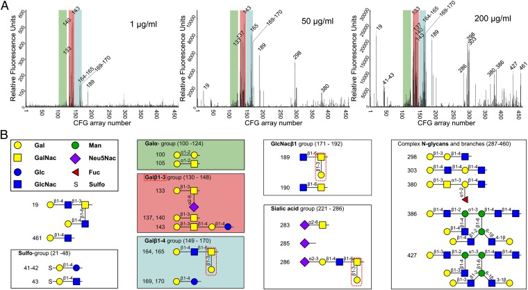
Glycan binding by the Epa1A domain. (A) Binding profiles of Epa1A at different protein concentrations (1, 50, and 200 μg/mL), using the CFG array V4.1 harboring 451 different glycan structures. Relative fluorescence units as monitored reflect relative affinities toward the corresponding glycan. Glycans bound by Epa1A are indicated by their CFG array numbers. Overall, Epa1A recognizes exclusively terminal galactosides. Green, Galα group; red, Galβ1–3 group; blue, Galβ1–4 group. (B) Groups that are recognized by Epa1A are shown; their locations within the CFG array are given in parentheses. Structural formulas are described according to the CFG nomenclature. At 200 μg/mL, Epa1A recognizes both Galβ1–3 (red) and Galβ1–4 (blue) glycans. However, the latter can harbor terminal β1–3 ramifications (red dotted lines). At low concentrations, Epa1A has a much narrower specificity profile.

