Fig. 5.
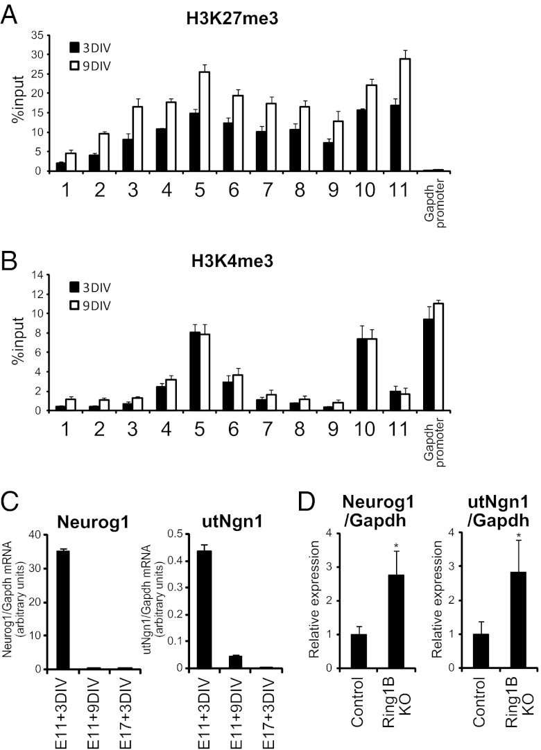
PcG proteins repress the expression of utNgn1 in the late stage of NPC development. (A and B) Primary NPCs isolated from the E11.5 neocortex were cultured for 3 or 9 DIV in suspension in the presence of FGF2 and EGF. Cells were then subjected to ChIP assays with antibodies to H3K27me3 (A) or to H3K4me3 (B) and with the PCR primers indicated in Fig. 1A. Data are means ± SEM from three independent experiments. (C) Primary NPCs isolated from the E11.5 or E17.5 neocortex were cultured for 3 or 9 DIV as in A, after which total RNA was isolated and subjected to qRT-PCR analysis of utNgn1 and Neurog1 mRNA. Data are normalized by the amount of Gapdh mRNA and are means ± SEM (n = 3). (D) Total RNA prepared from the neocortex of E18.5 Ring1bflox/flox; NestinERT2-Cre (Ring1B KO) and Ring1bflox/flox (control) embryos that were exposed to tamoxifen in utero at E12.5 were subjected to qRT-PCR analysis of Neurog1 mRNA and utNgn1. Data are normalized by the amount of Gapdh mRNA, are expressed relative to the corresponding value for control embryos, and are means ± SD from three embryos of each genotype. *P < 0.05 (Student t test).

