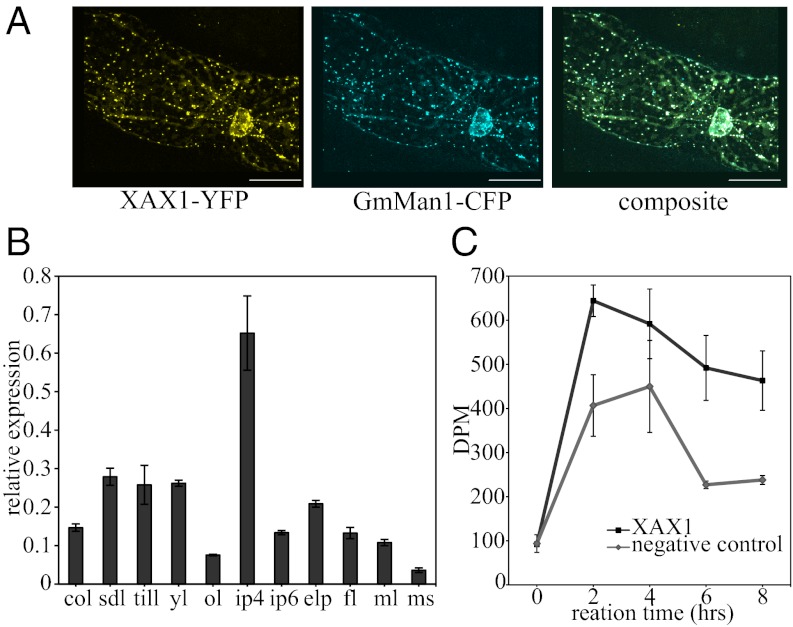
Fig. 3.
Characterization of XAX1 protein localization and function. (A) Subcellular localization of fluorescently tagged XAX1 proteins. Confocal imaging (40×) of onion epidermal cells expressing XAX1-YFP and GmMan1-CFP, the α-mannosidae Golgi marker. (Scale bar, 50 μm.) (B) Relative expression of XAXT in wild-type rice. col, coleoptile; sdl, 7 dpg seedling; till, 30 dpg tiller; yl, 30 dpg young leaf; ol, 30 dpg old leaf; ip4, immature panicle of 4 cm; ip6, developing panicle of 6 cm; elp, emerging panicle; fl, flag leaf; ml, mature leaf; ms, mature stem. (C) Xylosyltransferase activity reaction products. UDP-[14C]xylose incorporation to microsomes with an endogenous acceptor. Negative control is leaves infiltrated without XAX1 and represents the endogenous xylosyltransferase activity. Error bars are SEM with n = 3 biological replicates.

