Fig. 2.
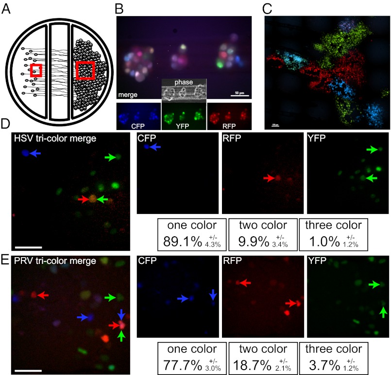
Three-color infection of compartmentalized neuronal cultures. (A) Schematic representation of compartmentalized neuronal culture system. Dissociated SCG cell bodies are plated in the far left soma (S) compartment and neurite extensions penetrate underneath two sequential barriers and extend into the far right neurite (N) compartment. Epithelial cells (Veros or PK15s for HSV and PRV, respectively) are plated onto the isolated neurites 1 d before neuron cell body infection. Red squares indicate the area imaged in B (Left box) and C (Right box). (B) SCG cells bodies in the S compartment were imaged 8 h after infection with a mixture of HSV-1 strain 17 that expresses mCereulean, EYFP, or mCherry. A merged image of all three fluorescent channels is presented with individual channels and phase-contrast image is inset below. (Scale bar, 50 µm.) (C) Vero detector cells plated on isolated neurites in the N compartment were imaged 48 h after neuronal cell body infection for fluorescence expression. A merged image of the three fluorescent channels is presented. (Scale bar, 100 µm.) (D) A representative image taken from time lapse imaging of the Vero detector cells demonstrating the spatially isolated initial infection of individual cells from isolated axons. Single-color, two-color, and three-color cells are marked as indicated. (Scale bar, 50 µm.) (E) A representative image taken from time lapse imaging of the PK15 detector cells labeled as described for D (Scale bar, 50 µm.)

